|
BASIC FACTS
ABOUT THE HUMAN BRAIN
We feel that in order to fully appreciate the seriousness of
sport-related concussions, or of any form of traumatic brain injury, it is
important to have a basic understanding of how the brain functions under
normal circumstances and what actually takes place when your brain suffers
an injury.
VIDEO SUPPLEMENT
Before you continue, it is recommended that you take the time
to watch a special 30 minute video that we have created entitled, Understanding
The Brain. By watching the video and reading the rest of this section,
you should have a good idea of what happens to your brain when it suffers
a traumatic brain injury.
THE
MOST COMPLEX ORGAN IN THE BODY
We
will now examine this incredible machine we call the brain. There is
nothing like the human brain. No man-made computer even comes close to the
capacity of the human brain. However, when the brain experiences a
traumatic injury, a whole lot of things happen that are cause for concern.
We will take a look at a very simple, basic explanation of how the brain
works under normal circumstances and what happens inside the brain when it
is injured.
As
you gain a better understanding of how your brain works, you will
appreciate why it is important for us to have an effective concussion
management program in place for student-athletes who suffer sport-related
traumatic brain injuries.
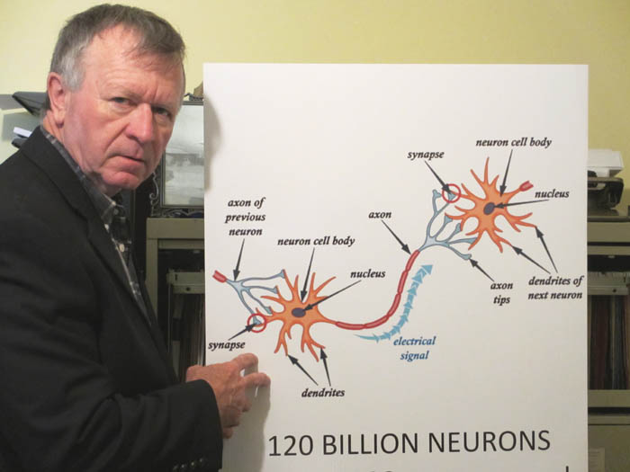
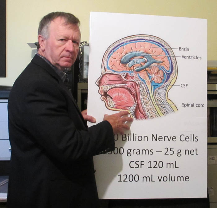 Robert
Kirwan is shown making the following presentation to a group of coaches
who were taking part in a Concussion Management Training Seminar. A
close-up photo of the skull is shown below. Robert
Kirwan is shown making the following presentation to a group of coaches
who were taking part in a Concussion Management Training Seminar. A
close-up photo of the skull is shown below.
The adult human
brain is a soft, jelly-like organ that weighs about 1500 grams (3 pounds)
and is about 1200 cubic centimeters in volume.
You could fit the human brain into one of the three
milk bags you get in a 4L package of milk.
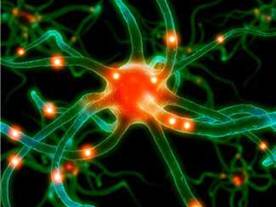
There are over 100 billion neurons in the brain. We
often refer to these as brain cells.
 Each of these neurons includes between 1000 and
10,000 protrusions called dendrites which are used to receive electrical
signals from other neurons. Each of these neurons includes between 1000 and
10,000 protrusions called dendrites which are used to receive electrical
signals from other neurons.
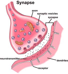
The electrical signals travel through axons,
which are long slender tubes and projections that conduct electrical
impulses and allow biochemical reactions to take place across a tiny space
called a synapse at the point where the axons meet up with dendrites.
Axons and dendrites don’t actually touch. They just come very close to
each other. Close enough for the chemical neurotransmitters to jump across
from the axons to receptacles in the dendrites.
Each neuron has one axon which takes electrical
impulses "from" the sending neuron to as many as 10,000
dendrites of other neurons.
The dendrites "receive" electrical
impulses from other neurons, then transform the energy to create its own
neural signal pattern before sending it to other neurons in its network
though its own axon.
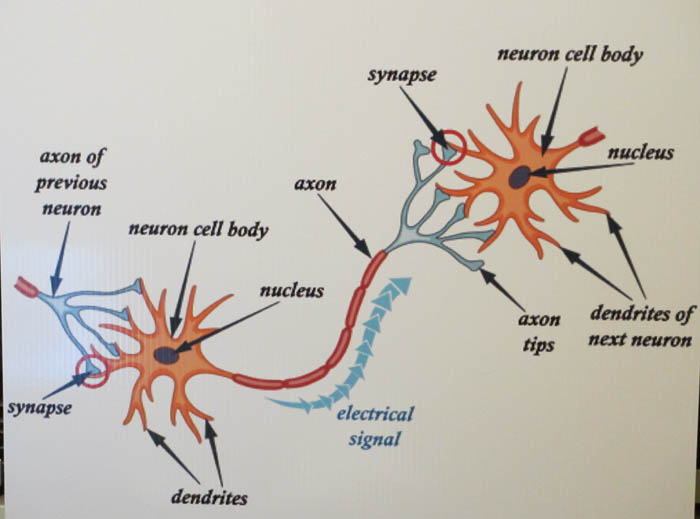
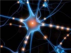
The diagram below will show you how the neurons
communicate with each other. Now imagine each axon branching off to go
throughout the brain, connecting to thousands of other neurons that will
become part of the specific communication network that is needed in order
for this particular function to take place. Imagine how many neurons will
be included in any one of these networks and you have some idea of just
how complex the operation of the brain really is.
To give you another idea of just how incredibly
small this complex structure is, if you could lay all of the axons that
are inside your brain connecting the nerve cells, end to end, you would be
able to go around the world at the equator over four times. That’s about
160,000 km of axons all jumbled up together inside your brain providing
the communication link between the 100 billion nerve cells contained in
your brain – the central nervous system.
All of this fits in a space about the size of a milk
bag and weighing about 3 lbs or 1500 grams.
The neurons and axons make up only part of the
volume of the brain. Scientists differ on just how much of the volume this
consists of, but the rest of the volume consists of glial cells. Glial
cells provide support and protection for the neurons and assist in some
way with the communication between neurons.
There are four main functions
of the glial cells.
-
They surround the neurons and axons, holding them in
place.
-
They insulate one neuron from another and keep the axons separated
from other axons so the wires don’t cross inadvertently.
-
They supply
nutrients and oxygen to the neurons.
-
And, they destroy and remove dead
neurons, essentially keeping the brain clean.
Early studies of the brain
estimated that up to 90% of the volume of the brain consists of glial
cells. More recent studies take the position that the balance is more like
50% of glial cells with the other 50% being neurons. Regardless, both
neurons and glial cells play critical roles in the central nervous system.
DEVELOPMENTAL DIFFERENCES BETWEEN ADULTS AND
CHILDREN
There are a number of theories why it takes a
child’s brain longer to recover from a concussion. The developmental
differences between an adult and an adolescent are significant and these
differences influence how the brain reacts to trauma.
The first consideration deals with the substance
which surrounds the axons. This substance is known as myelin. It is like
the plastic coating that you find on electrical wiring in your house. The
coating protects the wire and allows for efficient transmission of
electricity. You can twist and bend the wire and the coating protects the
copper wiring inside. The axons of an adult have the same kind of
protection. The myelin is built up and works to protect the axons from
injury. Concussions still occur in adults, but it takes more force to
damage the axons because of the protection from the myelin.
Children and adolescents have less myelin since
their brains are still developing. Therefore it is much easier for damage
to occur to the axons and it takes less force to cause stretching or
shearing of axons that are not as protected as with adults.
Another development issue has to do with the size of
the head which is disproportionately larger relative to body size during
childhood and adolescence. This extra size and weight influences the force
that is being applied to the brain as a result of blows received to the
head and body during sport competition.
The final development issue we will consider deals
with muscle development. The muscles in a child and adolescent are not yet
fully developed, therefore the student-athlete may not be strong enough to
brace for contact. This lack of development is critical in the neck area
which has a lot to do with the movement of the head following a body blow.
So when you consider the size and weight of the head
relative to the rest of the body; the lack of muscle strength; and the
lack of myelin protecting the
axons running through the brain, it is easy to see why children and
adolescents take longer to recover from concussions.
| COMMUNICATION SYSTEM BETWEEN NEURONS

|
 It is a very
complex process, but the ability of nerve cells to effectively communicate
with each other along a complicated network is what allows you to function
as a normal human being. It is a very
complex process, but the ability of nerve cells to effectively communicate
with each other along a complicated network is what allows you to function
as a normal human being.
A concussion changes the way the brain normally
functions which is why this is such a serious injury and should not be
taken lightly.
If you look to the diagram to the left, you will
notice that the axon from one neuron never actually touches the dendrite
of another neuron. Instead, it meets at a place that is called a synapse,
which is the name of the small space between the end of the axon and the
end of the dendrite. Let me repeat - the synapse is the name of the
"space" between the axon and the dendrite. This is an important
point to remember.
As amazing as it sounds, from what we know about the
brain, it would appear as if we have over 100 billion neurons, each with
up to 10,000 dendrites, connecting through a single axon to up to 10,000
other dendrites, and yet no two neurons are actually physically connected.
They are all separated by a small space at the synaptic junction.
The actual communication is by chemical
neurotransmitters that influence the receiving neuron. No two of the more
than 100 billion neurons are actually physically connected. This is an
amazing phenomenon that is hard to comprehend considering the small space
inside the skull.
So, to repeat, when an electrical signal is sent
through the axon, it creates a chemical reaction that produces
neurotransmitters which are sent across the synapse to receptors on the
dendrite. When this happens, the receiving neuron transforms the signal
from the sending neuron to its own special electrical signal and then
sends that signal along to thousands of other neurons through its own
axon.
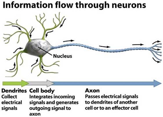
The diagram below will give you another overview of
how information flows through neurons throughout the brain.
AN INJURY INTERRUPTS THE FLOW OF INFORMATION
When your brain suffers an injury that results in a
concussion many things happen all at once and as a result some of those
dendrites and axons may be stretched or broken. There is also a tremendous
power surge as billions of neurons send out electrical impulses
simultaneously, releasing a cavalcade of neurochemicals from the axons in
the brain.
This results in a disruption or disconnection of the
pathways between many of the nerve cells and causes all kinds of problems
in the way messages are communicated and distributed throughout the brain.
With over 160,000 km of axons weaving their way through the brain to
neurons in many areas of the organ, the interruption of the signal pathway
along a single axon could have significant impact on the functioning of
the brain and may produce a wide variety of symptoms depending on which
pathways have been affected.
The power surge of energy as the neurons all fire up
their electrical signals at once, coupled with the release of chemicals
into areas of the brain where the chemicals may not have been before, adds
to the crisis situation and causes all kinds of unpredictable events to
occur.
THE BRAIN - A NEW FRONTIER
 Keep in mind that most of what we know about the brain has just recently been discovered. Keep in mind that most of what we know about the brain has just recently been discovered.
But what we do know for sure is that each one of the
100 billion nerve cells can connect with thousands of other nerve cells
through these dendrites and axons which wind their way around the brain.
In fact up until about the age of 20 your brain is
continually forming neural connections until you reach up to about 1,000
trillion connections between nerve cells. As you get older about half of
the connections are discontinued in a sort of pruning process, mainly
because they are not being used, but you will still end up with no less
than 500 trillion connections between neurons for most of your adult life.
The period when you have the greatest number of neural connections is
during adolescence, from ages 13 to 19, typically the years when you are
in the intermediate and senior grade levels of secondary school (Grades 7
through 12)
CENTRAL NERVOUS SYSTEM
 Dendrites and Axons, therefore, are similar to telephone wires or internet cables
carrying the messages being sent between nerve cells in the brain and
throughout the body via the spinal column to and from the brain. This is
why the brain is called the “central nervous system”. It acts a lot
like a bus terminal where signals are sent and then distributed elsewhere
depending on where they can be put to best use. Dendrites and Axons, therefore, are similar to telephone wires or internet cables
carrying the messages being sent between nerve cells in the brain and
throughout the body via the spinal column to and from the brain. This is
why the brain is called the “central nervous system”. It acts a lot
like a bus terminal where signals are sent and then distributed elsewhere
depending on where they can be put to best use.
Everything you do is the result of electrical
impulses and biochemical reactions that travel through some of the 160,000
km of axons connecting each of the 100 billion nerve cells in your brain
to thousands of other nerve cells, resulting in up to 1000 trillion
different connections in total, all producing chemical reactions across
the synapses that permit communication to take place.
As well, the neurons inside your brain are connected
through the brain stem and the spinal cord to the nerve cells and sensory
cells throughout your body, sending signals that tell your body how to
function.
Just reading these sentences involves thousands of
nerve cells being connected along hundreds of km of axons, producing
millions of neurotransmitters that are being taken in by millions of
receptors, and all of this happens in a split second. If I tell you to put
your finger on the letter Q on the key pad, just think of what your brain
has to go through to make your finger actually move to the keyboard
letter. This simple command requires memory, vision, muscle coordination,
reasoning, etc. All of this is instantaneous, even though the
communication is being sent along neural pathways that are in a variety of
different areas of the brain.
The brain is an incredible machine that is pretty
durable under normal circumstances. But if something happens to cause the
brain to suffer any kind of injury, there are so many things that can go
wrong because of its complexity.
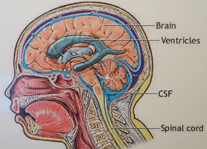
CEREBROSPINAL FLUID (CSF)
Something
else you need to know is that the brain is submerged in cerebrospinal
fluid (CSF).
This fluid occupies the open space inside the skull and among other
things, provides buoyancy for your brain.
CSF also protects the brain tissue from damage
against the inside of the skull during normal movement of the head or
body. It provides a cushion between the brain and the skull bone, so the
brain doesn't strike the skull very often under normal conditions.
CSF CONTROLS INTRACRANIAL PRESSURE
There is normally space for about 130 to 150 ml of CSF in side the skull
and it is replaced about 3 or 4 times a day, draining into the blood.
The intracranial pressure is maintained by the body at a fairly constant
level by maintaining just the right total volume of CSF; just the right
amount of blood flow to the brain; and obviously by the composition of the
brain itself.
Any increase or decrease in one of the three elements (CSF, blood flow,
or volume of the brain) means that one or both of the other two must be
reduced or increased in order to maintain the right amount of intracranial
pressure. Since the brain is a constant size and the blood flow doesn’t
change much, and since the CSF is constantly being produced and drained so
often each day, the body usually uses the amount of CSF production to keep
the pressure constant whenever the need arises.
HUGE IMPACT ON WEIGHT OF THE BRAIN
The cerebrospinal fluid provides buoyancy for the
brain, so even though the brain has an actual mass of about 1500 grams,
the net weight of the brain suspended in the normal amount of CSF is
equivalent to a mass of only 25 grams, or about the weight of two normal
sized grapes.
This is important since it allows the brain to
maintain its density without being impaired by its own weight which would
cut off blood supply and kill nerve cells in the lower sections of the
scull cavity without the right amount of CSF.
Keep
in mind that without the CSF the brain would feel 60 times heavier.
The amount of CSF is extremely important in order to
provide what is known as neutral buoyancy. This means that the net weight
of the brain allows it to be "suspended" in the CSF instead of
floating to the top of the skull or sinking to the bottom. The suspension
of the brain in this state of neutral buoyancy allows it to keep its shape
and density. If it sank or floated it would rest up against the top or
bottom of the skull, placing pressure on the blood vessels, restricting
blood flow and killing off neurons. The amount of CSF is critical to the
functionality of the brain.
Therefore,
as the brain is suspended inside the skull, it feels very light, which is
why we can move around a lot and not feel anything moving around in our
head. Even most rapid movements of the head would not produce much of an
impact against the side of the skull since the brain feels so light when
everything is normal.
RECAP…
So, to be clear, what you have inside your skull is your brain matter
(basically dendrites, axons, nerve cells) which takes up about 1200 ml of
space; the CSF fluid which takes up another 130 ml of space; and the
remaining portion consists of blood vessels. All of this is kept together
inside a bag called the dura.
|
PARTS
OF THE BRAIN AND THEIR FUNCTIONS
|
|
In order to better understand what happens when the brain is
injured, we would like to take a bit of time to examine the main
parts of the brain and their functions.
|
|
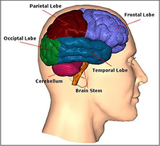 FRONTAL LOBES FRONTAL LOBES
The Frontal Lobes are located at the front part of your head, just
behind the forehead. This part of the brain is very prone to injury
because it is very close to the ridges of the skull and in many
instances with head-on force this area slams against the bone. This
part of your brain is responsible for helping you make plans,
organize things, solve problems, and effectively use your memory. It
is also the part of your brain that controls your emotions and
impulses and helps you maintain socially acceptable behaviour. It
also helps you with your ability to pay attention to details and to
make decisions. Finally, this area plays a huge role in your speech
and language abilities.
TEMPORAL LOBES
The temporal lobes are found at the sides of the brain behind the
frontal lobes right around the level of your ears. This part of the
brain is responsible for your hearing and for helping you to
recognize and understand sounds and speech and also to produce
speech for communication purposes.
OCCIPITAL LOBES
This part of your brain is located right at the lower back of the
head and is where you process visual information which is sent from
your eyes. It helps you make sense out of what you see and perceive
shapes, colours, sizes, and distance.
PARIETAL LOBES
This part of your brain is located right behind your frontal lobes.
It is the part of your brain that integrates the sensory information
that comes from all parts of your body when you touch things or feel
hot, cold, etc. The parietal lobes also help you with some of your
balance and give you the ability to navigate around without bumping
into things.
CEREBELLUM
The cerebellum is located at the back of the brain and controls your
balance, movement and co-ordination. It allows you to perform the
physical activities that are necessary for sports and just for
movement in general. It is the area of your brain that is most
involved in coordination of all parts of your body.
BRAIN STEM
The brain stem is located at the base of the brain and controls all
of the functions that are necessary for survival, such as your
breathing, heart rate, and blood pressure. These are all of the
involuntary functions of the brain that you do without thinking.
A COMPLEX SYSTEM
The brain is a very complex system that serves us well normally.
However, when brain trauma occurs that results in a concussion, the
damage can be widespread and can impact any number of these
sections. Because of the interconnection of neurons, and the fact
that each neuron can be connected to up to 10,000 other neurons, and
each of those neurons can be connected to up to another 10,000
neurons, and so on, it is safe to say that whatever happens to one
neuron may in fact have an effect that reaches all parts of the
brain. We will accept that in most cases the impact may be
negligible, but nonetheless, there is an impact and if enough
neurons are damaged or enough of the axons are stretched and/or
sheared, there can be significant and widespread damage.
You
don’t need a medical degree to see that the different parts of the
brain work together in order for one to function normally. Damage to
the Frontal Lobes will definitely have an effect on how you respond
to what you see and the signals coming from your Occipital Lobes.
And if you have damage to your Cerebellum, thus affecting your
balance, it is going to have an impact on multiple regions of your
brain.
This
is why any force to the body that results in the brain moving
violently inside the skull gives cause for concern. Let us see what
happens to the brain when it is injured.
|
|
WHAT
HAPPENS TO THE BRAIN WHEN IT IS INJURED?
|
WHAT IS A CONCUSSION?
There seems to be general agreement that a concussion is caused by a
direct blow to the head, face, neck or any other part of the body. Loss of
consciousness is not necessary for a concussion to occur. In fact, only a
small percentage of concussions involve loss of consciousness.
The force of this contact, no matter where it occurs, causes the brain to
move violently from side to side, front to back or rotationally within the
skull. As a result, the brain as a whole is stretched or squashed slightly
as it bangs against the inside of the skull, causing it to change its
shape and become temporarily deformed. It very quickly returns to its
original shape, even though it may be a bit swollen from striking the
inside wall of the skull.
No matter what definition you use, the fact remains that a concussion
changes the way the brain functions. What is not known at this time is how
long or how permanent the damage will remain.
Many people refer to a concussion as a "temporary Traumatic Brain
Injury" or a temporary TBI. You will often see the definition include
reference to the "rapid onset of short-lived impairment of
neurological function that resolves spontaneously".
However, there is great debate going on now as research points that the
impairment of neurological function may not repair as rapidly as once
thought and the resolution may not be as spontaneous as we had hoped.
This temporary impairment may be true for the most obvious symptoms such
as headache and dizziness, but the long-term impact of a concussion may
result in impairment of emotional and psychological functions as a result
of the changes that occur in the brain.
In fact, there are studies that have found middle aged adults who
suffered concussions while in college exhibiting premature brain aging and
deficiencies in concentration, balance and motor control many years after
suffering their concussions. It is most likely that most people who are
suffering from these kinds of functional deficits may simply attribute
them to normal aging and getting older, not even relating any symptom or
deficit to their history of concussions. And yet, there may be things they
could have done during rehabilitation that might have reduced or
eliminated these functional deficits, thus impacting on their quality of
life many years after the injury. Our goal in developing the most
effective student-athlete concussion management program possible is to
reduce the long-term consequences of sport-related concussions.
A
CONCUSSION IS A PROCESS – NOT AN EVENT
Evidence is being produced by researchers which proves clearly that a
"concussion is a process". It is not an event. And this process
does not simply involve "healing and recovery". Many symptoms of
concussion do not present themselves for hours, days, weeks or months. In
fact some people admit to experiencing concussion-like symptoms for many
years following an injury.
We will concede that there may well be a rapid onset of short-lived
impairment of neurological function in some areas that resolve
spontaneously, but what about the long-term impairment that does not
resolve. What about personality changes? What about anxiety and mood
disorders? What about interpersonal relationship skills? What about one's
attitude towards life? These are all recognized as signs and symptoms of
concussion but they are also unfortunately accepted by most people as part
of growing up and normal development. They may not be that normal after
all.
Admittedly, we all change our personality slightly from time to time. We
all have periodic bouts of anxiety and we are all moody from time to time.
We all have some difficulties with relationships and our attitude towards
life is often affected by our environment and the people around us. But
for young people who suffer a concussion, are these changes part of their
natural evolution, or are they consequences of their brain injury? And is
there something we can do to reduce the risk of life-altering
consequences?
Symptoms of a concussion may also not be evident until you are required
to perform a specific task. For example, you may not even know that you
are no longer able to recall math facts until you are asked to recite your
times table. You may not realize that you get dizzy riding a bike until
you have a chance to ride a bike. You may not know you have problems
adjusting your vision when things are being thrown quickly in your
direction from the side until this actually happens. These symptoms take
time to present themselves and they will only be noticed if you have
people around you who are looking for signs and symptoms of concussion.
That is why we use the "partner approach" to concussion
management.
WHAT HAPPENS DURING A CONCUSSION?
Axons get their shape from internal structures called microtubules which
look like a string of sausages strung together. As the shape of the brain
gets temporarily deformed from the twisting or rapid acceleration and/or
deceleration, the axons may stretch or break. Normally, since your brain
is constantly jiggling like a mold of jello, axons are often stretched
gently with no damage to any of the internal skeletal structure that is
found inside axons. This is what is often referred to as a “slow
stretch”.
If the axons are stretched too quickly, they tend to stiffen up causing
their internal skeletons to become destroyed and the axons will shear,
causing a total interruption of signals. In most cases, concussions are
less severe injuries where the axons do not actually shear, but rather are
stretched with enough force that they don’t quite rip apart but still
sustain significant damage to their internal skeletal structure.
For example, if the axon is stretched hard enough, the microtubules that
act like conveyor belts carrying nutrients from one end of the axon to the
synaptic connections in its network may break at some point. When this
“conveyor belt” is broken, the supplies that are being carried will
continue to flow but they will basically “fall off” at the break and
will collect inside the axon. This causes a “bulb” to form inside the
axon. More importantly, it prevents the part of the axon beyond the break
from receiving the nourishment and supplies it needs to survive.
Eventually, the part of the axon that is not receiving nourishment will
wither away and die, thus disconnecting from the original axon. That means
that signals that would normally have gone along that axon will no longer
get through. This then causes the axons with the bulbs of protein to also
shrivel up and die because they can no longer do what they are supposed to
do and the neuron will die as well. All communication that was conducted
that one neuron will then cease.
There are some injuries where the damage is beyond repair, but the
communication is still continuing in a faulty manner. The signals are
getting through but they are not clear. In this case the damaged
connection may end up corrupting the entire system with static
communication.
Dr. Douglas Smith of the
University
of
Pennsylvania
and a number of his colleagues have done extensive research on concussions
and axonal damage. What they found is that if you stretch an axon gently
the first time, it produces an increase in the number of tiny pores that
line the outside skin of an axon. These pores allow sodium and calcium to
come inside. If you stretch the axon gently a second time shortly after
the first time, these tiny pores became enlarged and sodium and calcium
came rushing in. Other scientists had previously discovered that increased
levels of calcium in an axon created an enzyme that actually ate away the
internal structure of the axon. Therefore, the implication is that if a
person suffers a seemingly minor blow to the head or body, there may not
be any obvious symptoms of concussion present, but the stretched axons
will be extremely vulnerable if there is another minor blow. That is why
some people are surprised when they receive serious concussion-like
symptoms from what seemed like a very small force. It’s because the
axons were vulnerable at the time from the stretching caused by the first
blow.
“THE
METABOLIC CASCADE”
When
the brain suffers from a force as a result to a blow to the head or some
other part of the body, it experiences a "power surge" producing
an extreme amount of chemical neurotransmitters, effectively
"lighting" up the entire brain with electrical charges. This
surge only lasts a minutely brief period of time and seems like a
mini-seizure. The physical movement causes neurons and axons all over the
brain to be pulled, twisted and stretched.
The
neurons send out signals through the axons to allow sodium and calcium to
enter through the tiny pores on the outer skin that have been enlarged by
the twisting and stretching. At the same time potassium is allowed to rush
out of the neurons through the axon openings. The problem with too much
sodium is that it also brings in water which can cause swelling of the
axons and thus dangerously increase intracranial pressure. Calcium
produces an enzyme that eats away at the internal structure of the axons.
Once
the initial power surge is over, the brain immediately attempts to restore
the equilibrium and get things back to normal levels. The first thing the
neurons do is send a signal to pump potassium back into the axon and pump
sodium back out. The potassium counteracts the effects of sodium by
neutralizing its electrical charge. This process requires a lot of energy
which is usually produced inside the neuron by something called the
mitochondria, which acts like an internal power plant for each cell.
The
mitochondria require fuel in the form of glucose to produce energy.
Glucose is carried to the neurons by the blood flow in the brain. The
demand on the cell for energy causes a drain on the supply which causes
the brain to lose power and operate on a slower speed. The brain then
demands for an increase in blood flow in order to bring in more glucose to
the mitochondria to repair the damaged areas. However, the message somehow
is disrupted and the blood flow to the brain is actually slowed down. No
matter how many signals the neurons send out for more fuel, there is no
increase in blood flow and the cells are in danger of dying. Because of
this the brain releases high quantities of potassium in order to try to
calm things down even more.
Since
each
dendrite or axon may be part of a communication line that carries impulses
to thousands of nerve cells as it winds its way around the brain, any
damage to a dendrite or an axon can impact many areas of the brain in the
network other than just the area where the original damage was caused.
This domino affect can cause symptoms that may seem unusual based on the
point of impact, but neurons in one part of the brain connect to neurons
in other parts of the brain and may be part of a communication link with
many other functions.
This is why we often see a variety of symptoms when a person suffers a
concussion. The damage can affect your cognitive, physical, emotional and
psychological functioning and it can play havoc with your sleep patterns
and relationships.
|
THE HEALING PROCESS IN
THE BRAIN
|
AUTOMATIC RESPONSE
When the brain experiences a trauma, the body goes into an automatic
emergency protection mode and a number of things take place that are
designed to help the brain begin the healing process. However, it is this
healing process that may actually put the student athlete in jeopardy if
the proper procedures are not followed when an injury occurs.
REDUCTION IN BLOOD FLOW TO THE BRAIN CAUSES AN ENERGY CRISIS
Immediately
following a brain injury where there is damage to nerve cells, dendrites
and axons, along with some swelling of the brain, there is an automatic
response by the body that results in a reduction of blood flow to the
brain. While this may reduce internal bleeding if a blood vessel breaks,
it also means that the damaged area of the brain is being deprived of
oxygen and energy that it needs in order for healing to take place. This
"energy crisis" makes the stretched or torn dendrites, axons and
damaged neurons (nerve cells) extremely vulnerable and seriously impedes
the healing process. In fact, studies have shown that a large number of
neurons can die during this initial period because of the lack of oxygen
and energy that result from the reduced blood flow. Death of a neuron is
permanent.
REDUCTION OF CSF LEVELS
INCREASES WEIGHT OF THE BRAIN
Because
of the swelling that generally occurs in the damaged area of the brain,
the intracranial pressure may begin to rise slightly. In order
to compensate for this dangerous increase in pressure the body reduces the
amount of CSF present around the brain since this is the quickest way for
the body to naturally reduce intracranial pressure. The brain simply
drains out some CSF and does not replace it until the pressure is back to
normal.
While this is happening, the reduction in CSF has a critical impact on
the buoyancy of the brain. There isn’t as much CSF surrounding the brain
as there is under normal conditions, therefore the net weight of the brain
feels much heavier than the usual 25 g. Remember that the brain itself
would weigh about 1500 grams (3 pounds) without the CSF. With the normal
amount of CSF it would only weight 25 g because it is suspended in the
fluid. This buoyancy effect is the reason why you seem to weigh less when
you are swimming.
SUSCEPTIBLE TO FURTHER INJURY
This reduction of blood flow and CSF is going on in your head, even as
you are coming back to the bench to “shake it off” and recover from
your immediate symptoms. The emergency response in your brain is going
into overdrive and you may not even be aware of what is happening unless
you begin to feel a bit of a headache or a bit dizzy.
Keep in mind that studies have shown that in up to 80% of the cases where
a student-athlete has suffered a concussion, the student-athlete was not
aware of any symptoms right away. So this could be taking place without
you having any knowledge that you were injured in the first place. The
headaches and dizziness may come minutes or hours after the injury.
With less buoyancy causing the brain to feel much heavier after an
original injury, it is extremely susceptible to serious injury if the body
suffers another blow and the brain suffers an additional trauma. Even a
minor, seemingly insignificant blow to the body could result in a much
more serious injury than the original blow because the much heavier brain
will be hitting the inside of the skull and twisting with much more force
because of the increased net weight.
On top of this, because of the original injury, the damaged axons have
been stretched and become brittle. If there is another trauma that
triggers an immediate surge in chemicals and electrical impulses through
these stretched and brittle pathways, the pressure may cause the stretched
and weakened axons to break completely and this will completely interrupt
communication along those pathways.
COMPLETE SHUT-DOWN IS NECESSARY
This is why we strongly suggest that a student-athlete who has suffered
what appears to be a serious blow that could have resulted in concussion
should remain out of action for at least the rest of that day and reduce
both physical and cognitive exertion until we can be sure of the extent of
the damage.
Everything may seem fine on the surface and there may be no indication of
obvious symptoms of a concussion immediately after the event, but inside
the skull the body may have already taken necessary precautions as part of
its emergency response, thus leaving the brain exposed to further and
potentially much more serious damage.
DANGER OF REPEAT CONCUSSION
This is why a “second repeat concussion” is often more severe than
the original concussion. The original trauma may have stretched and
damaged the axons and brain cells, but they may not have been completely
broken. This means that even if their function has been reduced, they have
not been discontinued. They can still operate in a reduced capacity and
gradually they will return to their original condition and regain their
flexibility. Eventually the flow of chemicals and electrical impulses will
be able to reach their pre-injury levels and everything should be back to
normal within a period of time.
On the other hand, if you don’t allow the proper time for healing and
you don’t try to avoid overextending the damaged areas, you are taking a
chance that the lines will burst, and then you are in serious trouble.
There is no guarantee that you will ever regain full functioning in these
areas if they are damaged a second, third or subsequent time.
What is even more frightening is that you could damage those injured
areas simply by increasing the electrical and chemical impulses by
watching television, playing video games, texting on the cell phone, or
listening to music. You don't just need to worry about physical exertion.
You also have to be concerned about cognitive exertion. You need to shut
down all physical activity and you also must shut down your brain!
|
CONSEQUENCES
OF A CONCUSSION
|
DYSFUNCTION – TEMPORARY OR PERMANENT?
Most experts agree that about 80% of people who suffer a concussion
appear to be symptom-free within 10 days to two weeks of getting the
injury. However, and this is an extremely important point to remember,
especially with our student-athletes, there is no consensus about whether
subtle changes remain in the brain following those 10 days. Furthermore,
we need to be especially concerned about the 20% of people whose symptoms
do not go away within the first ten days. What is happening to their
brains as they wait for recovery? What must we do to help them cope with
what they are going through?
Therefore, when we speak of a student-athlete who has a concussion, we
mean that the student-athlete is experiencing a complex process that is
affecting the normal functioning of a part of his brain that may have an
impact on many areas of his life. Our goal is to do everything in our
power come up with a rehabilitation program that will make this truly one
of those temporary conditions and prevent it from having life-altering
consequences.
MANY SYMPTOMS DO NOT SHOW UP IMMEDIATELY
What many people fail to understand is that some of the symptoms may last
much longer than others, and as we are going to find out, many of the
symptoms of concussion do not produce obvious signs. In fact, many of the
symptoms only show up much later and often as a result of a second blow to
the body that transmits a force to the same area of the brain that was
injured in the first place. This is why CMP will always take the position
that once any sign, symptom or behavior consistent with concussion is
observed or experienced, you must assume that there are other symptoms
that you may not yet be aware of.
We all know that many student-athletes experience a competitive event
where they are “dazed” and have their “bell rung”. After a couple
of minutes of rest they may be able to “shake it off” and feel ready
to go back into action. This temporary symptom may have resolved itself in
a few minutes, but that doesn’t mean that the brain is totally
recovered.
For example, symptoms such as headache, nausea, dizziness, vision
problems, vomiting, loss of balance, confusion, feeling in a fog, ringing
in the ears, and slurred speech may be evident and temporary. In fact they
may appear and then disappear within minutes.
However, other behavioural symptoms may only be noticed over time, often
over days or weeks. For example, decreased playing ability may have
resulted from the injury, but those signs may not be evident right away,
especially if the player is removed from play. Mood disorders, such as
sadness, anxiety, irritability, aggressiveness and other inappropriate
emotions may appear as subtle changes that are hardly noticeable at first
and which may simply be passed off as normal reactions to being injured
and out of action.
Cognitive signs may only be noticed when the student-athlete returns to
the classroom or may only be noticed by parents/guardians during normal
day-to-day activities. Being slower to react when responding to questions
an having difficulty concentrating or remembering information are symptoms
of serious symptoms that are on-going and which may take some time to
resolve.
Sleep difficulties may only be noticed by parents/guardians and can
easily be overlooked or passed off as other problems. A student-athlete
who complains about being drowsy may seem normal unless it is about being
more drowsy than usual. A parent will notice if his/her child is having
trouble falling asleep or if he/she is sleeping more or less than usual.
These are all signs of concussion symptoms that cannot be ignored.
Since a concussion is actually a “dysfunctioning of the brain” that
is the result of a force to the head, even though the student-athlete may
feel he has recovered physically, the impact of the blow may still be
creating problems emotionally, intellectually and psychologically.
SUSCEPTIBLE TO REPEAT CONCUSSIONS
In fact, the number of people who seem to be more susceptible to repeat
concussions once they suffer the first one gives rise to the theory that
even once symptoms seem to be gone, there are still unseen vulnerabilities
that may place the person at risk. In fact, the area of the brain that was
originally damaged may end up being more vulnerable to future damage or
the area may have weakened surrounding areas that end up becoming more
vulnerable. The thing is - we just don't know enough about the brain to be
certain. However, based on what we do know about the
brain it is not surprising to find out that once you receive the first
concussion it is much easier to get repeat concussions is absolutely true.
SUBCONCUSSIONS
Experts also believe that many student-athletes may suffer what is
referred to as subconcussions.
These are very minor injuries that do not produce any obvious symptoms,
but over time if a person suffers enough repetitive subconcussions, the
accumulative deterioration of the nerve cells and axons cause long-term
changes in brain function that often appear in mid-life and have a
significant effect on behaviour and personality.
Subconcussions may also weaken enough areas of the brain so that a full
concussion is inevitable with the right amount of force. Since
subconcussions are almost impossible to detect in that they produce no
obvious symptoms, we should adopt the philosophy that if it is felt that a
student-athlete suffered a hit to the body or head that "might
have" produced enough force to the brain to cause a concussion, it
very likely resulted in at least a subconcussion and warrants further
investigation and monitoring.
Despite the fact that many experts believe that symptoms from a
concussion are temporary, there is no doubt that as the recovery process
unfolds the brain is extremely vulnerable to further trauma which may
result in serious long-lasting consequences that go far beyond what we
would call temporary. Therefore, the question remains: is a subconcussion
a concussion? Are signs and symptoms necessary in order for the brain to
be experiencing a concussion? Is a subconcussion simply a minor
concussion? Can subconcussions be responsible for post-concussion
symptoms? In fact, can a person have post-concussion symptoms without even
being aware that he/she suffered a concussion in the first place? If
he/she suffered a subconcussion instead?
The reality is that most adults have suffered from some traumatic brain
injury at some point in their life. The injury may have come while playing
sports or an accident. And anyone who has played a contact sport surely
has suffered some degree of a concussion at some point in their playing
career. So when a person claims to have never suffered a concussion it may
just be that they were not able to identify the signs and symptoms of a
concussion or that they had what we now call subconcussions where signs
and symptoms were not obvious.
POST-CONCUSSION SYMPTOMS
Statistics show that at least 10% of individuals with a concussion suffer
post-concussion symptoms for months and years, especially if they were not
properly treated after a concussion. And many others may have functional
deficits that they do not relate to previous concussions and/or
subconcussions, but nonetheless they do exist.
What we do know from research studies is that well after they have
"recovered" from an injury, student-athletes who have suffered
two or more concussions are more likely to report having concussion-like
symptoms such as headaches, balance problems, sensitivity to light and
noise, trouble concentrating and sleeping, irritability and nervousness
than those student-athletes who only experienced one concussion or none.
Student-athletes with two or more concussions have also been found to be
more likely to score lower on measures of attention and concentration and
tend to do worse in school than those with one or no concussions. All of
this points to the importance of having a solid concussion management
program in place that will make sure student-athletes fully recover from
each concussion before being allowed to return to play.
IMPACT OF DAMAGE TO THE FRONTAL CORTEX
Researchers are learning more and more about the brain every year. They
have now found evidence that the Frontal Cortex or as they are often
called, the frontal lobes of the brain seems to be the most common region
of injury from a concussion. Damage to this part of the brain can cause a
wide variety of symptoms since the neurons found in the frontal cortex are
involved in motor function, problem solving, spontaneity, memory,
language, initiation, judgment, impulse control, and social and sexual
behaviour. This is considered our emotional centre and is where we exhibit
our personality.
Frontal lobe damage has been associated with reduced
ability to perform fine motor movements and diminished strength in the
arms, hands and fingers. Difficulty in speaking has also been common with
this type of injury.
It has also been noted from studies that even when a
student-athlete appears to have recovered completely from a concussion,
there is evidence of a lingering interference with attention and memory,
both which would impact tremendously on the ability of a student-athlete
to handle the demands being made in the classroom.
So when we discuss the temporary nature of
concussions or we talk about concussions completely healing, we cannot
ignore the changes in social behaviour or personality that often follow a
concussion. We tend to pass these changes off as part of growing up, or
simply changes that were triggered by the injury, however, researchers may
eventually find evidence that concussions actually change the course of a
person's life and thus have permanent repercussions.
We must avoid the tendency to diminish the
consequences of a concussion by stating that it is a mild traumatic brain
injury that will resolve spontaneously. The explosion of neurotransmitters
during the power surge in the brain at the time of impact may in fact
result in permanent changes to the neural pathways and the synaptic
architecture of various regions of the brain, such as the frontal cortex
which is connected to just about every other area of the brain. The
reorganization and rerouting of the neural pathways may bring a
student-athlete to close proximity with pre-injury functioning, but
changes may still exist and in fact the person may need to strengthen
those reconfigured pathways all over again.
INJURY THRESHOLD
Adding to the mystery surrounding concussions is the fact that studies of
athletes have shown that the amount of force and the location of the
impact are not necessarily correlated to the severity of the concussion or
its symptoms. This has lead to some confusion among experts about the
amount of force that is actually required in order to cause a concussion.
Studies have also found that concussions occur over a wide range of
impact magnitudes and that individuals have different levels of
biomechanical concussion thresholds. A blow of a certain level of
intensity that gives one person a concussion may not have the same affect
another.
Furthermore, it has also been found that the injury threshold
“within” an individual is dynamic and not at all constant. This means
that a certain magnitude of impact will produce different results in an
individual depending on the level of impact tolerance that person has at
the time of impact. It changes with the day and the time of day.
There is a school of thought that if the injury tolerance is indeed
dynamic in an individual, then this tolerance threshold may be influenced
by the number of subconcussive impacts sustained by the athlete in the weeks or months prior to the impact that causes the
concussion. Or that the longer a player participates in a sport, the more
likely he is going to be concussed at some point in time because of the
cumulative effect of subconcussive impacts. This will receive further
study over the next number of years, but when you think of what happens to
the pathways when they stretch after a trauma, and if you imagine these
pathways going through the stretching and healing process a number of
times, it makes sense that after a certain amount of stretching they would
become weaker. The more often you stretch a balloon for example, the
weaker it gets and eventually it will break.
It must never be forgotten that a concussion can alter the brain’s
physiology for anywhere from hours to weeks, setting in motion a variety
of events that interfere with the functioning of the neurons in the brain.
The damage that occurs in most affected brain cells is usually reversed,
but a few cells may die after the injury and some cells may take longer to
heal than others. This is just something normal to expect.
NOW
YOU HAVE A BETTER APPRECIATION OF WHAT IS AT STAKE
By
now you should now have a much better appreciation of what is at stake
when it comes to managing concussions that are sustained by
student-athletes. Every year we are increasing our knowledge base about
the brain and how it works. Unfortunately, much of what we are learning is
pointing out the errors we have made in the past when it came to dealing
with sport-related head injuries. The challenge facing all of us today is
to move forward, not in fear, but with care, choosing to implement
protocols and procedures that err on the side of caution. We can no longer
ignore the fact that any damage to the brain may produce life-altering
consequences, changing the entire course of a person’s life.
|